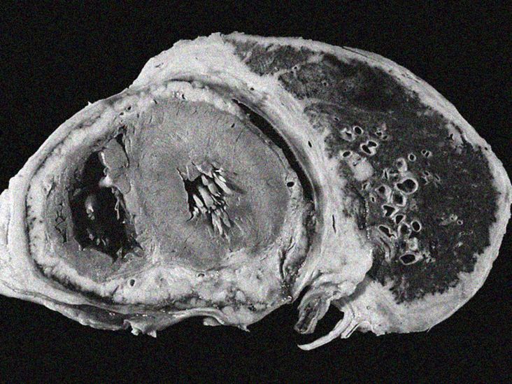
What does lung cancer look like on an X-ray, CT scan, or PET scan? It sounds like a straightforward question, but medical imaging rarely behaves like a police lineup where one suspicious spot steps forward and confesses. Lung cancer images can show masses, nodules, fluid, collapsed lung tissue, enlarged lymph nodes, or subtler changes. However, infections, scars, inflammation, and noncancerous growths can create similar pictures.
That is why doctors do not diagnose lung cancer by staring dramatically at one glowing screen. They combine imaging findings with symptoms, previous scans, smoking and exposure history, laboratory results, andmost importantlya tissue biopsy when cancer is suspected. This guide explains what doctors may see, what the major imaging tests can and cannot reveal, and what patients commonly experience during the diagnostic process.
What Does Lung Cancer Look Like on Medical Images?
There is no single appearance shared by every lung cancer. A tumor may appear as a small round nodule near the outer edge of a lung, an irregular mass close to a major airway, or a hazy area that is difficult to distinguish from inflammation. Its appearance depends on its size, location, growth pattern, cancer type, and whether it has affected nearby structures.
Common findings that may prompt further testing include:
- A new pulmonary nodule or mass
- A nodule that has grown since an earlier scan
- Irregular, lobulated, or spiky borders
- A partly solid or ground-glass opacity
- Enlarged lymph nodes in the center of the chest
- Airway obstruction or collapse of part of a lung
- Fluid around the lung, known as a pleural effusion
- Possible spread to the bones, liver, adrenal glands, brain, or other organs
None of these findings proves that cancer is present. Most lung nodules found on CT scans are not malignant. Old infections, granulomas, scar tissue, autoimmune disease, and benign tumors can all produce suspicious-looking spots. In other words, radiology occasionally gives harmless objects the lighting of a movie villain.
Lung Cancer on a Chest X-Ray
How an X-ray Creates the Image
A chest X-ray sends a small amount of radiation through the chest. Air absorbs little radiation, so healthy air-filled lungs generally look dark. Bones, the heart, blood vessels, fluid, and dense tissue absorb more radiation and appear lighter.
A lung tumor may therefore appear as a white or gray area within the darker lung. It might look like a rounded nodule, a larger mass, an area of lung collapse, or an indirect change caused by blocked airflow.
What an X-ray May Show
Chest X-rays may reveal relatively large tumors, especially when they are surrounded by air-filled lung. They can also show pneumonia, fluid around a lung, enlarged structures near the center of the chest, or a section of lung that is not expanding normally.
A central tumor may obstruct a bronchus and cause tissue behind the blockage to collapse or become repeatedly infected. In such cases, the X-ray may show the consequences of the obstruction more clearly than the tumor itself.
Why Chest X-Rays Can Miss Lung Cancer
An X-ray compresses three-dimensional anatomy into a two-dimensional picture. Ribs, blood vessels, the heart, diaphragm, and other structures overlap. Small nodules can hide behind these structures, while tumors near the center or top of the chest may be especially difficult to recognize.
For this reason, a normal chest X-ray does not completely rule out lung cancer when symptoms or risk factors remain concerning. Chest X-rays are also not recommended as the primary screening test for people at high risk. Low-dose CT is more sensitive for detecting small lung nodules and is the recommended screening method.
CT Images of Lung Cancer
Why CT Scans Provide More Detail
Computed tomography uses X-rays and computer processing to create thin cross-sectional images, often described as slices. Instead of seeing the entire chest layered into one picture, a radiologist can examine the lungs section by section and reconstruct images in multiple planes.
CT scans can identify nodules that are too small or poorly positioned to be visible on a standard X-ray. They also provide information about a finding’s exact dimensions, shape, density, borders, and relationship to airways, blood vessels, lymph nodes, and the chest wall.
Common CT Appearances
A suspicious lung lesion may appear as a solid nodule, a partly solid nodule, a ground-glass opacity, or a larger mass. Ground-glass opacity is a hazy area through which underlying vessels may still be visible. It can occur with infection or inflammation, but persistent ground-glass or part-solid nodules sometimes represent early lung adenocarcinoma.
Radiologists also study the edges of a lesion. Smooth, stable nodules are often less concerning than growing lesions with irregular or spiculated borders. “Spiculated” means thin lines extend outward from the nodule, rather like an unfriendly little sunburst. It raises concern but is not a diagnosis by itself.
Other factors include:
- Whether the nodule contains fat or calcium
- How quickly it changes between scans
- Whether it forms a cavity
- Its location within the lung
- The presence of additional nodules
- Associated lymph-node enlargement
- The patient’s age, exposures, and cancer history
Contrast-Enhanced CT
Some diagnostic CT scans use intravenous contrast material to make blood vessels and certain tissues easier to evaluate. Contrast may help doctors examine the relationship between a mass and nearby vessels, identify enlarged lymph nodes, and assess the liver, adrenal glands, or other possible sites of spread.
Low-dose screening CT, by comparison, is usually performed without contrast. It is designed to find lung nodules in people who do not have symptoms but have an elevated risk of developing lung cancer.
Low-Dose CT Screening Images
Screening and diagnostic testing are not quite the same. Screening looks for disease in people who have no symptoms. A diagnostic scan investigates a symptom or an abnormality already found on another test.
The U.S. Preventive Services Task Force recommends annual low-dose CT screening for adults ages 50 through 80 who have at least a 20 pack-year smoking history and currently smoke or quit within the previous 15 years. Screening generally stops when a person has not smoked for 15 years or develops a health condition that substantially limits life expectancy or the ability to undergo curative lung surgery. Eligibility and insurance rules can differ, so patients should confirm the current criteria with a healthcare professional and insurer.
A pack-year equals smoking an average of one pack per day for one year. For example, one pack daily for 20 years and two packs daily for 10 years both equal 20 pack-years.
The scan itself usually takes only a few minutes. The patient lies on a table, briefly holds their breath, and moves through a short, open scanner. There are no needles or tunnels worthy of an action-movie escape scene, and screening LDCT normally requires no contrast injection.
What a Positive Screening Result Means
A positive LDCT result does not automatically mean lung cancer. It means the scan found something that meets criteria for additional evaluation. Depending on the nodule’s size and appearance, the next step may be another CT after a defined interval, a diagnostic CT, PET/CT, consultation with a lung specialist, or biopsy.
Small, low-risk nodules are often monitored rather than immediately biopsied. Comparing new images with earlier studies is particularly valuable. A nodule that has remained unchanged for a long period may be less concerning than one that is clearly growing.
PET/CT Images and Metabolic Activity
Positron emission tomography adds functional information to anatomical imaging. Before a typical cancer PET scan, a patient receives a small amount of a radioactive glucose-like tracer, commonly fluorodeoxyglucose, or FDG. Tissues that absorb more tracer appear brighter on the images.
Because many cancer cells consume glucose rapidly, lung tumors and metastases may show increased FDG uptake. Combining PET with CT helps doctors match an area of metabolic activity to its precise anatomical location. PET/CT is frequently used to evaluate whether lung cancer may have reached lymph nodes or distant organs and to help plan treatment.
Bright Does Not Always Mean Cancer
Infection and inflammation can also absorb FDG and appear bright. Recent surgery, radiation therapy, granulomatous disease, and active pneumonia may produce false-positive findings. Conversely, some small or slow-growing cancers may show little tracer uptake.
PET/CT therefore adds an important piece to the puzzle but does not replace biopsy. It can estimate how suspicious a lesion is and reveal useful targets for tissue sampling, but it cannot reliably identify the exact cancer subtype by itself.
MRI Images in Lung Cancer Care
Magnetic resonance imaging uses a strong magnetic field and radio waves rather than ionizing radiation. MRI is not usually the first test for finding a small lung nodule because moving air-filled lungs are generally evaluated better with CT.
However, MRI can be extremely useful when doctors need detailed images of the brain, spinal cord, nerves, chest wall, or structures near the top of a lung. A brain MRI may be performed during staging because lung cancer can spread to the brain, sometimes before neurological symptoms appear.
MRI may also help evaluate whether a tumor has extended into nearby soft tissues. Whether it is needed depends on the cancer type, stage, symptoms, and planned treatment.
Ultrasound and Other Imaging Procedures
Ultrasound is not normally used to search through air-filled lung tissue because sound waves do not travel well through air. It can, however, identify fluid around the lung and guide thoracentesis, a procedure that removes pleural fluid for testing.
Ultrasound may also be used during bronchoscopy. Endobronchial ultrasound allows a specialist to view lymph nodes and other structures next to the airways and guide a needle into them. This can provide tissue needed for diagnosis and staging without open surgery.
CT guidance is another common approach. During a CT-guided needle biopsy, imaging helps the physician place a needle through the chest wall and into a suspicious nodule or mass.
Can Imaging Tell Which Type of Lung Cancer Is Present?
Imaging may suggest certain possibilities, but it cannot reliably determine whether a tumor is adenocarcinoma, squamous cell carcinoma, small cell lung cancer, or another subtype. Some cancers favor particular locations or growth patterns, yet these features overlap too much for a definitive visual diagnosis.
A biopsy provides cells or tissue for examination by a pathologist. Laboratory testing can identify the cancer type and may check for biomarkers or genetic changes that influence treatment. This distinction is critical because surgery, radiation, chemotherapy, immunotherapy, and targeted therapy are selected according to the tumor’s pathology, stage, molecular characteristics, and the patient’s overall condition.
How Doctors Use Images to Stage Lung Cancer
Staging describes how far cancer has spread. CT, PET/CT, MRI, and tissue sampling may all contribute. For non-small cell lung cancer, doctors evaluate the primary tumor, nearby lymph nodes, and distant metastases. This is commonly summarized through the TNM system:
- T: Size and local extent of the main tumor
- N: Involvement of regional lymph nodes
- M: Spread to distant parts of the body
Imaging may reveal enlarged or metabolically active lymph nodes, but size and brightness are imperfect clues. A normal-sized node can contain cancer, while an enlarged node may be reacting to infection. That is why suspicious lymph nodes are often sampled through endobronchial ultrasound, mediastinoscopy, or another biopsy technique.
Imaging also helps determine whether a tumor can be surgically removed, define radiation treatment fields, select a safe biopsy route, and measure treatment response.
Understanding Common Words in a Lung Imaging Report
Nodule
A small rounded or irregular opacity within the lung. A nodule is a description, not a cancer diagnosis.
Mass
A larger abnormal area. A mass generally requires prompt evaluation, although noncancerous causes remain possible.
Ground-Glass Opacity
A hazy area that does not completely obscure the structures beneath it. Causes range from temporary infection to early cancer.
Spiculation
Thin strands extending from a lesion into surrounding tissue. Spiculation may increase suspicion for malignancy.
Atelectasis
Partial collapse or incomplete expansion of lung tissue. It may occur when a tumor blocks an airway, but mucus and other conditions can also cause it.
Hilar or Mediastinal Adenopathy
Enlarged lymph nodes near the central lungs or within the mediastinum, the space between the lungs.
Pleural Effusion
Fluid between the lung and chest wall. Cancer is one possible cause among many, so fluid analysis may be necessary.
Stable
No meaningful change compared with an earlier scan. Stability can be reassuring, although the necessary monitoring period depends on the lesion’s type and clinical context.
Images That Resemble Lung Cancer
Several conditions can imitate lung cancer on radiographs or CT scans:
- Pneumonia and other infections
- Tuberculosis or fungal granulomas
- Scar tissue from an old infection or injury
- Inflammatory and autoimmune disorders
- Benign hamartomas
- Rounded atelectasis
- Pulmonary abscesses
- Vascular abnormalities
Radiologists use features such as growth, density, calcification pattern, border shape, and comparison with earlier scans to narrow the possibilities. Even so, some findings remain indeterminate. Medicine occasionally has to admit, “That spot needs another look,” rather than manufacture certainty where none exists.
Common Experiences During the Lung Imaging Journey
For many patients, the most difficult part is not the scan. It is the waiting. An X-ray may be completed in minutes, yet the phrase “abnormal opacity” can occupy every available corner of the brain until the follow-up appointment.
A common journey begins with a persistent cough, shortness of breath, chest discomfort, unexplained weight loss, or an unrelated medical visit. Someone may receive an X-ray for pneumonia, a preoperative evaluation, or even an injury, only to learn that the radiologist noticed a lung nodule. Others enter the process through annual low-dose CT screening while feeling completely healthy.
The first emotional reaction is often to assume the worst. That is understandable, but a nodule is not synonymous with cancer. Many patients are surprised to learn how frequently scars and old infections appear on lung images. Asking the clinician to explain the nodule’s size, density, borders, and comparison with previous scans can replace at least some of the uncertainty with useful facts.
Patients also describe confusion when one test leads to another. An X-ray may lead to CT because CT provides greater detail. CT may lead to PET/CT because doctors want to evaluate metabolic activity and possible spread. PET/CT may lead to biopsy because only tissue can establish the diagnosis. This sequence does not necessarily mean the situation is steadily worsening. Each test answers a different question.
The practical details can help reduce anxiety. A chest CT is painless and usually quick. Patients may be asked to hold their breath for several seconds. If intravenous contrast is used, it can create a temporary warm sensation or metallic taste. Before a PET scan, patients typically receive preparation instructions, rest quietly after the tracer injection, and spend more time at the imaging center than they would for a routine CT.
Biopsy days often feel more serious because they move the process from “What does the image suggest?” to “What are these cells?” Depending on the location of the abnormality, tissue may be collected through bronchoscopy, a needle guided by CT, lymph-node sampling, or surgery. Patients should ask how the biopsy will be performed, what complications are possible, whether enough tissue will be collected for molecular testing, and when results are expected.
Another common experience is discovering that scan reports sound much scarier than the doctor’s explanation. Radiologists deliberately document possibilities and recommend follow-up when uncertainty exists. Words such as “suspicious,” “indeterminate,” or “cannot exclude malignancy” communicate risk; they do not announce a final diagnosis.
Keeping copies of imaging reports and making sure specialists can access the actual image files may prevent unnecessary repetition. Earlier studies are especially helpful because growth over time is one of the most important clues. A tiny spot that has not changed across years tells a different story from one that has doubled in size.
Some people find it useful to bring a trusted person to appointments. A second pair of ears can record measurements, next steps, and timelines while the patient’s brain is temporarily busy playing every alarming medical drama it has ever seen.
Useful questions include:
- What exactly did the scan show?
- How large is the nodule or mass?
- Was it present on an earlier image?
- Which features are reassuring or concerning?
- Is another CT appropriate, or is a biopsy needed?
- What will the next test tell us that this one cannot?
- Who will coordinate the follow-up?
The experience may involve uncertainty, but it should not involve silence. A clear follow-up planwith a specific test, responsible clinician, and expected time frameis an important part of safe care.
Conclusion
Lung cancer images can provide crucial clues, but no single image tells the entire story. Chest X-rays may reveal larger abnormalities, CT scans show detailed anatomy, PET/CT evaluates metabolic activity and possible spread, and MRI examines areas such as the brain and nearby soft tissues. Ultrasound and CT can also guide procedures that collect the tissue needed for a definitive diagnosis.
The most important lesson is that an abnormal image is the beginning of an evaluation, not its conclusion. Many lung nodules are benign, and even highly suspicious findings must be interpreted in clinical context. Prompt follow-up, comparison with previous images, specialist review, and appropriate biopsy help turn an unsettling picture into an informed plan.