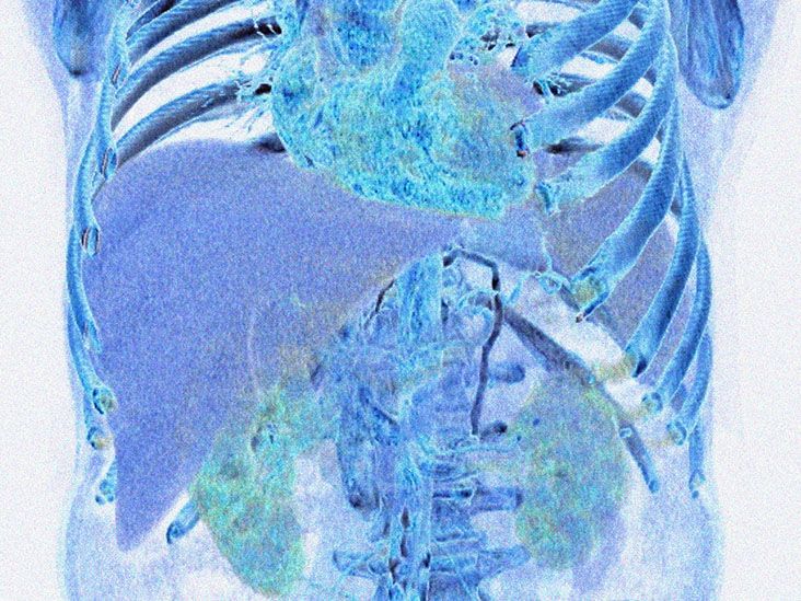
The portal vein does not receive much attention when everything is working properly. Yet this large blood vessel performs a crucial job: It carries nutrient-rich blood from the intestines, stomach, spleen, gallbladder, and pancreas to the liver. When a blood clot partially or completely blocks that route, the condition is called portal vein thrombosis, or PVT.
Some people with portal vein thrombosis have sudden abdominal pain. Others develop complications gradually, and many have no obvious symptoms when the clot is first discovered. That unpredictability is one reason PVT can be difficult to recognize without imaging.
Treatment also is not one-size-fits-all. Doctors consider when the clot formed, how much of the vein is blocked, whether nearby veins are involved, the presence of cirrhosis or cancer, and the person’s bleeding risk. In other words, treating PVT is less like following a cake recipe and more like solving a medical puzzle with several moving pieces.
What Is Portal Vein Thrombosis?
Portal vein thrombosis occurs when a thrombus, commonly called a blood clot, develops inside the portal vein or one of its major branches. The obstruction may be partial, leaving some room for blood to pass, or complete.
The clot may also extend into connected vessels such as the splenic vein or superior mesenteric vein. Extension into the mesenteric circulation is especially concerning because those veins drain the intestines. Severe obstruction can reduce intestinal blood flow and cause bowel ischemia or tissue death.
Recent Versus Chronic PVT
A recent or acute portal vein thrombosis has developed relatively recently, although the exact age of a clot is not always obvious. It may cause abdominal pain, nausea, fever, or no symptoms at all.
With chronic PVT, the body may create a network of small collateral blood vessels around the blocked portal vein. This process is called cavernous transformation or a portal cavernoma. These new vessels act like improvised side streets around a closed highway, but they do not always carry blood efficiently. The resulting pressure can contribute to an enlarged spleen, low platelet counts, and fragile varices in the stomach or esophagus.
Bland Thrombus Versus Tumor Thrombus
Doctors must also distinguish a conventional blood clot, known as a bland thrombus, from tumor growing into the portal vein. Tumor thrombus may occur with hepatocellular carcinoma and certain other abdominal cancers. This distinction matters because anticoagulants can help treat a blood clot, but they do not remove cancerous tissue invading a vein.
Portal Vein Thrombosis Symptoms
Portal vein thrombosis symptoms vary widely. A small partial clot may be found incidentally during an ultrasound, CT scan, or MRI performed for another reason. More extensive thrombosis is more likely to cause noticeable problems.
Possible Symptoms of Recent PVT
- Abdominal pain, often in the upper abdomen
- Abdominal tenderness or bloating
- Nausea or vomiting
- Diarrhea
- Fever, particularly when an abdominal infection is involved
- Reduced appetite
- New or worsening fluid buildup in the abdomen
Abdominal pain is common in symptomatic acute PVT, but pain alone cannot confirm the diagnosis. Gallstones, pancreatitis, ulcers, appendicitis, and approximately half the contents of a medical textbook can also cause abdominal discomfort. Imaging is therefore essential.
Symptoms of Chronic PVT and Portal Hypertension
Chronic obstruction can increase pressure within the portal venous system. Signs and symptoms may include:
- An enlarged spleen
- A low platelet count
- Abdominal swelling from ascites
- Enlarged veins in the esophagus or stomach
- Vomiting blood
- Black, tarry, or bloody stools
- Anemia, fatigue, or shortness of breath from gastrointestinal bleeding
- Worsening jaundice or confusion in someone with liver disease
In a person with cirrhosis, newly developed PVT may appear as worsening liver function, increased ascites, or variceal bleeding rather than a dramatic new pain.
When PVT Is an Emergency
Seek emergency care for severe or persistent abdominal pain, especially when accompanied by repeated vomiting, abdominal rigidity, bloody diarrhea, fever, fainting, confusion, or low blood pressure. These symptoms may indicate intestinal ischemia, major bleeding, sepsis, or another life-threatening complication.
Vomiting bright red blood or material resembling coffee grounds is also an emergency. The same applies to black, sticky stools, which may signal bleeding from esophageal or gastric varices.
What Causes Portal Vein Thrombosis?
PVT often results from a combination of sluggish blood flow, damage or inflammation near the vein, and a greater tendency for blood to clot. These factors resemble the classic ingredients of venous thrombosis elsewhere in the body, but the underlying conditions are frequently related to the liver or abdomen.
Cirrhosis and Chronic Liver Disease
Cirrhosis is one of the most important portal vein thrombosis risk factors. Scar tissue increases resistance to blood flow through the liver, slowing circulation within the portal vein. At the same time, liver disease can create complicated changes in both clotting and bleeding mechanisms.
Having cirrhosis does not mean a person is naturally “auto-anticoagulated.” Someone may have abnormal clotting tests and still develop a significant blood clot. This apparent contradiction is one reason treatment decisions require a hepatologist or another clinician experienced in liver-related coagulation disorders.
Cancer
Liver cancer, pancreatic cancer, stomach cancer, and other abdominal malignancies may increase the likelihood of PVT. Cancer can promote clotting throughout the body, compress the portal vein, or directly invade it.
Imaging specialists examine whether the material inside the vein enhances with contrast, expands the vessel, or connects to a tumor. These features may help distinguish tumor thrombus from a noncancerous clot.
Inherited and Acquired Clotting Disorders
Portal vein thrombosis can be associated with conditions that make blood more likely to clot, including:
- Factor V Leiden
- Prothrombin gene mutations
- Antiphospholipid syndrome
- Protein C, protein S, or antithrombin deficiency
- Myeloproliferative neoplasms
- Paroxysmal nocturnal hemoglobinuria
Doctors do not automatically order every available thrombophilia test for every patient. Testing is more likely when PVT occurs without cirrhosis or cancer, appears at a young age, recurs, affects multiple vascular areas, or is accompanied by a suggestive personal or family history.
Inflammation, Infection, Surgery, and Trauma
Inflammation near the portal vein can activate clotting. Potential triggers include acute pancreatitis, inflammatory bowel disease, diverticulitis, appendicitis, gallbladder infection, and other intra-abdominal infections.
When an infected clot develops in the portal system, the condition is called pylephlebitis or septic portal vein thrombosis. Fever, abdominal pain, and bloodstream infection may occur. Treatment usually includes antibiotics and, in selected cases, anticoagulation.
PVT may also develop after abdominal surgery, liver transplantation, splenectomy, bariatric surgery, pancreatic procedures, or significant abdominal trauma.
Pregnancy, Hormones, and Childhood Risk Factors
Pregnancy and estrogen-containing medications can increase clotting tendency, particularly when another risk factor is present. In infants and children, PVT may be associated with severe dehydration, sepsis, an infected umbilical stump, congenital portal vein abnormalities, or underlying liver disease.
How Is Portal Vein Thrombosis Diagnosed?
A clinician begins by reviewing symptoms, medical history, medications, previous blood clots, liver disease, recent surgery, cancer history, and family history. A physical examination may reveal abdominal tenderness, ascites, jaundice, an enlarged spleen, or signs of gastrointestinal bleeding.
Blood Tests
Blood testing may include a complete blood count, liver function tests, kidney function, and coagulation studies. A low platelet count may result from an enlarged spleen. Liver test results can be normal in people without liver disease, so normal bloodwork does not rule out PVT.
Doppler Ultrasound
Doppler ultrasound is commonly the first imaging test. It can show material blocking the portal vein and evaluate the speed and direction of blood flow. It is noninvasive and does not expose the patient to ionizing radiation.
Ultrasound quality can be affected by body anatomy, intestinal gas, operator experience, and the location of the clot. An unclear or positive examination may therefore be followed by cross-sectional imaging.
CT and MRI
Contrast-enhanced CT or MRI can confirm the diagnosis, measure how much of the vein is blocked, and determine whether the clot extends into the mesenteric or splenic veins. These scans can also look for bowel injury, inflammation, cirrhosis, or an abdominal tumor.
In patients with cirrhosis, detailed imaging is especially useful for distinguishing bland PVT from tumor invasion and for planning possible liver transplantation or interventional treatment.
Endoscopy
An upper endoscopy may be recommended when chronic PVT or cirrhosis raises concern about esophageal or gastric varices. During this procedure, a clinician examines the esophagus and stomach for enlarged veins that could bleed.
Portal Vein Thrombosis Treatment
The goals of portal vein thrombosis treatment are to prevent clot extension, restore blood flow when possible, protect the intestines, manage the underlying cause, and reduce complications of portal hypertension.
Anticoagulant Medication
Anticoagulants are commonly used for recent non-tumoral PVT, particularly in people without cirrhosis. These medications do not instantly dissolve a clot. Instead, they limit further clotting while the body gradually breaks down existing thrombus.
Options may include low-molecular-weight heparin, warfarin, or a direct oral anticoagulant. The best choice depends on liver and kidney function, pregnancy status, cancer treatment, possible drug interactions, bleeding risk, insurance coverage, and the amount of clinical evidence available for the patient’s situation.
For noncirrhotic PVT caused by a temporary risk factor, treatment may continue for at least six months after the trigger has been corrected. Longer or indefinite anticoagulation may be recommended for recurrent thrombosis, persistent clotting disorders, active cancer, or other continuing risks.
Never begin, stop, or adjust an anticoagulant without medical supervision. These medications can prevent serious complications, but they can also cause dangerous bleeding when used incorrectly.
Treatment in People With Cirrhosis
Management is more individualized when cirrhosis is present. A recent clot blocking less than half of the main portal vein or limited to smaller intrahepatic branches may sometimes be observed with imaging approximately every three months.
Anticoagulation is more likely to be considered when a recent clot blocks more than half of the main portal vein, involves the mesenteric vessels, continues to grow, affects more than one vascular bed, or could complicate liver transplantation.
For chronic, completely occlusive PVT with cavernous transformation, anticoagulation may provide little opportunity to reopen the original vein. Care may instead focus on preventing variceal bleeding and treating other consequences of portal hypertension.
Antibiotics for Infection
Pylephlebitis requires antimicrobial treatment directed at organisms commonly found in the digestive tract. Blood cultures, imaging, and treatment of the original infection are important. An abscess or another infected source may require drainage or surgery.
Thrombolysis, Thrombectomy, and TIPS
Procedures are reserved for selected patients because they carry meaningful bleeding and procedural risks. Catheter-directed thrombolysis uses clot-dissolving medication delivered near the obstruction. Mechanical thrombectomy physically removes or disrupts thrombus.
A transjugular intrahepatic portosystemic shunt, commonly called TIPS, creates a channel through the liver to redirect blood flow. Portal vein recanalization combined with TIPS may be considered when PVT occurs with refractory ascites, recurrent variceal bleeding, or a need to improve portal vein access before liver transplantation.
Suspected bowel ischemia requires urgent involvement of hepatology, hematology, interventional radiology, gastroenterology, and surgery. Waiting for symptoms to “see how they feel tomorrow” is not a sound strategy when intestinal blood flow may be threatened.
Treatment of Tumor Thrombus
When cancer has invaded the portal vein, treatment centers on the malignancy and may include systemic therapy, radiation-based approaches, locoregional liver treatment, surgery, or supportive care. Anticoagulation may still be used when a separate bland clot is also present, but it does not treat tumor tissue itself.
Possible Complications
Complications depend on the clot’s location, extent, duration, and underlying cause. They may include:
- Portal hypertension
- Esophageal, gastric, or intestinal varices
- Life-threatening gastrointestinal bleeding
- Ascites
- Splenomegaly and low platelet counts
- Portal biliopathy involving the bile ducts
- Intestinal ischemia or bowel infarction
- Sepsis from an infected portal vein clot
- Technical difficulties during liver transplantation
Recovery and Outlook
The outlook for portal vein thrombosis is strongly influenced by the underlying illness. A recent noncirrhotic clot treated promptly may partially or completely recanalize. Chronic PVT may remain blocked while collateral vessels maintain adequate circulation.
Follow-up imaging helps determine whether the clot is shrinking, stable, or extending. Patients taking anticoagulants also need monitoring for bleeding, medication interactions, and changes in liver or kidney function.
People with cirrhosis, cancer, an inherited clotting condition, or previous recurrent thrombosis usually require longer-term specialist follow-up. The clot may be only one chapter of the medical story rather than the entire book.
Can Portal Vein Thrombosis Be Prevented?
Not every case is preventable, but risk may be reduced by treating liver disease, cancer, pancreatitis, and abdominal infections promptly. Hospital teams may use preventive anticoagulation when appropriate after surgery or during serious illness.
Patients should tell their clinicians about prior blood clots, clotting disorders, pregnancy, hormone use, cancer, and upcoming procedures. Those prescribed anticoagulants should take them consistently and report unusual bruising, persistent nosebleeds, blood in urine, black stools, or other possible bleeding symptoms.
General wellness habits support liver and cardiovascular health but cannot replace condition-specific care. Avoiding heavy alcohol use, maintaining a healthy weight, managing metabolic disease, and obtaining treatment for viral hepatitis may help reduce liver damage that contributes to portal hypertension.
What the Portal Vein Thrombosis Experience Can Feel Like
The following is a patient-centered composite based on commonly reported clinical experiences. It does not describe one identifiable person and should not be used to predict an individual outcome.
For some patients, the experience begins with a surprise. A person undergoes an abdominal ultrasound to investigate mildly abnormal liver tests, and the report unexpectedly mentions “possible portal vein thrombosis.” The phrase sounds alarming, particularly because most people have never heard of the portal vein before that afternoon. A follow-up CT scan is ordered, and suddenly the calendar fills with appointments involving gastroenterology, hematology, and perhaps hepatology.
The first challenge is often uncertainty. Is the clot new or old? Is it related to cirrhosis, inflammation, cancer, or a clotting disorder? Does it involve the mesenteric veins? Will anticoagulation be necessary? Patients may hear several unfamiliar termsrecanalization, cavernous transformation, varices, thrombophiliabefore they have fully absorbed the original diagnosis.
Someone with acute symptoms may have a very different beginning. Abdominal pain grows steadily over several days and is accompanied by nausea or fever. Because the pain is vague, the person initially assumes it is indigestion or a stomach bug. When the discomfort becomes persistent, imaging reveals a portal vein clot. Treatment may start in the hospital with injections or an intravenous anticoagulant before transitioning to medication taken at home.
Anticoagulation brings relief and a new collection of practical concerns. Patients may wonder whether they can exercise, travel, drink alcohol, undergo dental work, or take over-the-counter pain relievers. They may become hyperaware of every bruise. A small purple mark that once would have been ignored can suddenly receive the investigative attention of a crime scene.
Clear instructions help. Patients generally benefit from knowing which medication interactions matter, what bleeding signs require urgent care, and whom to contact before a procedure. A written medication list can prevent confusion when several specialists are involved.
For a person with chronic PVT, the experience may revolve less around the clot itself and more around portal hypertension. Follow-up can include blood counts, liver tests, repeated imaging, and endoscopy to look for varices. If varices are found, preventive medication or endoscopic banding may be discussed. The absence of abdominal pain does not necessarily mean follow-up is optional.
Emotional responses vary. Some patients become anxious about the clot extending. Others are frustrated when doctors cannot identify a single cause. An “unprovoked” diagnosis may feel unsatisfying, but medicine does not always provide a neat villain wearing a name tag.
Recovery often happens gradually. A three-month scan may show that the clot has shrunk but not disappeared. Another scan may be needed. Treatment decisions are revisited as the clinical picture changes. For transplant candidates, maintaining or restoring portal vein flow may have special importance because extensive thrombosis can make surgery more complicated.
The most useful experience is usually a coordinated one. Patients do better when they understand the plan: why anticoagulation is or is not being used, when imaging will be repeated, how portal hypertension will be monitored, and what symptoms require emergency care. PVT may be uncommon, but a clear roadmap makes it considerably less mysterious.
Conclusion
Portal vein thrombosis is a blood clot affecting a major vessel that delivers blood to the liver. It may produce sudden abdominal pain, cause gradual complications of portal hypertension, or remain silent until discovered on imaging.
Cirrhosis, cancer, abdominal inflammation, surgery, infection, pregnancy, and clotting disorders are among the possible causes. Doppler ultrasound, CT, and MRI help determine the clot’s location, extent, age, and relationship to nearby organs or tumors.
Treatment may involve anticoagulants, antibiotics, management of varices, or specialized procedures such as portal vein recanalization and TIPS. The safest approach depends on the individualnot merely on the presence of a clot. Prompt evaluation is especially important when severe pain, intestinal symptoms, gastrointestinal bleeding, fever, or confusion develops.