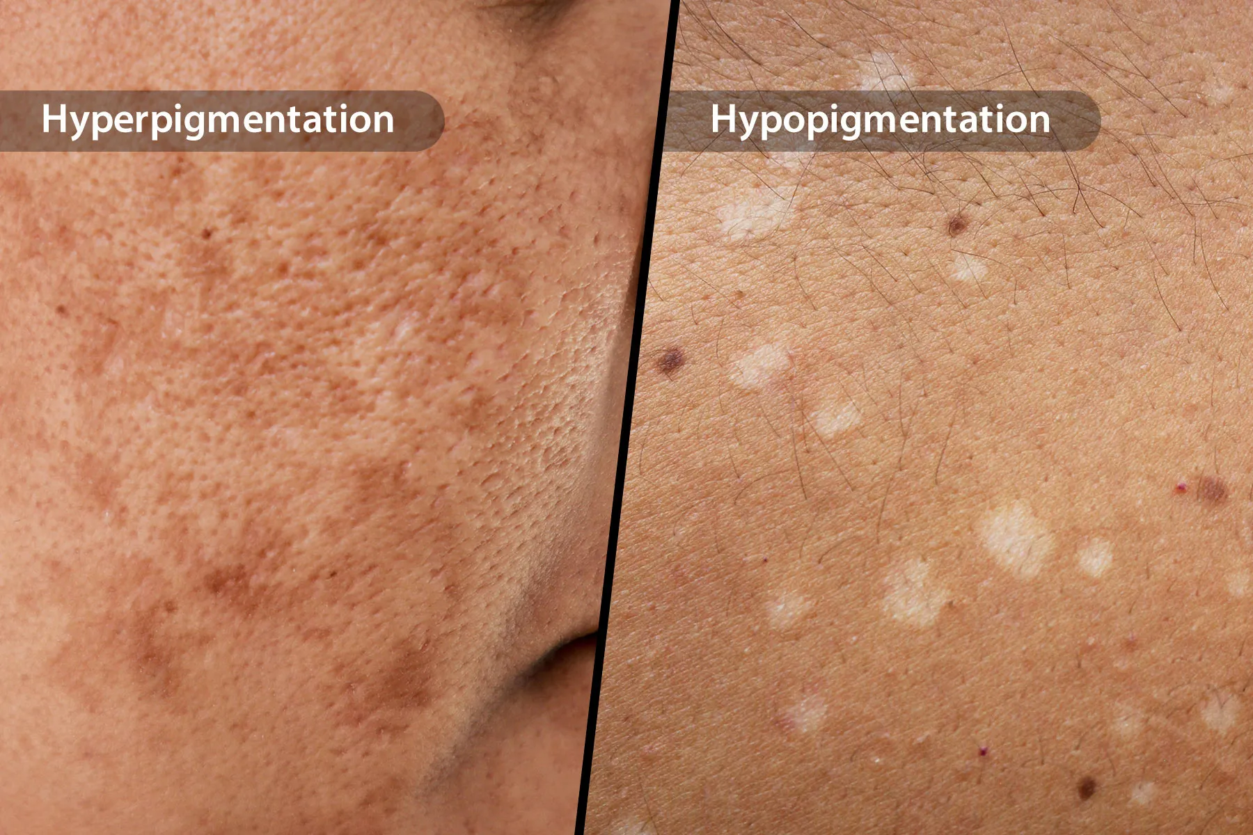
Skin color is wonderfully complicated. It can deepen after a sunny vacation, fade after a rash, darken during pregnancy, or develop patches that seem to appear without sending a calendar invitation. Two common terms used to describe these changes are hyperpigmentation and hypopigmentation.
Hyperpigmentation describes skin that becomes darker than the surrounding area, while hypopigmentation means an area has less pigment and looks lighter. When pigment is completely absent, dermatologists often use the more specific term depigmentation. These changes are usually cosmetic, but they can occasionally point to an infection, autoimmune disease, medication reaction, hormonal condition, or another health issue.
How Skin Pigmentation Works
Much of your skin color comes from melanin, a natural pigment produced by cells called melanocytes. These cells live mainly in the epidermis, the outer layer of the skin. They package melanin into tiny structures and distribute it to neighboring skin cells, helping determine the color of your skin, hair, and eyes.
Melanin also helps absorb ultraviolet radiation. However, having more melanin does not provide complete protection from sunburn, premature aging, or skin cancer. Every skin tone benefits from sun protection.
Pigment changes can occur when melanocytes produce too much melanin, make too little, become damaged, disappear from an area, or distribute pigment unevenly. Sometimes the amount of melanin is normal, but another substance, such as a medication or its breakdown products, becomes deposited in the skin and changes its appearance.
What Is Hyperpigmentation?
Hyperpigmentation is the darkening of an area compared with a person’s usual skin tone. Depending on skin color and the depth of the pigment, a patch may appear tan, brown, dark brown, gray, blue-gray, reddish brown, or nearly black. It may affect a tiny spot, several patches, or a large portion of the body.
Hyperpigmentation itself is not a diagnosis. It is a visible result with many possible causesrather like a “check engine” light for pigment, although often far less dramatic.
Post-Inflammatory Hyperpigmentation
Post-inflammatory hyperpigmentation, commonly shortened to PIH, develops after inflammation or injury. Acne, eczema, psoriasis, insect bites, burns, cuts, allergic reactions, and aggressive cosmetic treatments can all leave darker marks after the original problem improves.
PIH is especially common and often more noticeable in medium-to-deep skin tones. The discoloration may fade naturally, but improvement can take months or, in some cases, years. Picking pimples, scratching rashes, and constantly switching between harsh products may keep the inflammatory cycle running. In other words, the skin remembers drama longer than anyone asked it to.
Melasma
Melasma causes blotchy areas of darker pigmentation, most commonly on the cheeks, forehead, nose, upper lip, or jawline. Hormonal changes, genetic susceptibility, ultraviolet radiation, and visible light can contribute. Melasma frequently appears during pregnancy or while using certain hormonal medications, although it can affect people of any sex.
Melasma is persistent and may return after successful treatment, particularly when sun and visible-light protection are inconsistent. It is not contagious or cancerous, but it can have a significant emotional impact.
Sun-Related Dark Spots
Freckles and solar lentiginesoften called sun spots or age spotsdevelop after cumulative ultraviolet exposure. They tend to appear on the face, shoulders, upper chest, forearms, and backs of the hands. Although many are harmless, a new or changing dark spot should not automatically be assigned the cheerful label “just sun damage.”
Other Causes of Hyperpigmentation
Darkening may also be associated with friction, medication use, contact reactions, genetic conditions, nutritional problems, or systemic illnesses. Diffuse bronzing, especially when accompanied by fatigue, weight loss, dizziness, vomiting, or salt cravings, needs medical evaluation because adrenal disorders are among the possible causes.
Some medicines can stimulate melanocytes or leave deposits in the skin. The pattern and color depend on the medication, dose, duration of use, sun exposure, and individual susceptibility. Never stop a prescribed drug solely because pigmentation appears; contact the prescriber first.
What Is Hypopigmentation?
Hypopigmentation occurs when an area contains less pigment than the surrounding skin. The patches may look off-white, pale pink, cream-colored, or simply lighter than the person’s baseline complexion. Hypopigmentation is different from depigmentation, in which pigment is absent and the skin may appear distinctly white.
Light spots are often easier to notice after surrounding skin tans. That does not necessarily mean the condition suddenly worsened; the contrast may simply have become stronger.
Post-Inflammatory Hypopigmentation
Inflammation can sometimes reduce pigment instead of increasing it. Eczema, psoriasis, burns, infections, cosmetic procedures, chemical exposure, and skin injuries may temporarily disrupt melanocyte activity. As the skin heals, color often returns gradually, although deeper injury or scarring may cause lasting changes.
Vitiligo
Vitiligo is a chronic autoimmune disorder in which the immune system attacks melanocytes. It causes smooth white patches that may enlarge or develop in new areas. Common locations include the face, hands, feet, elbows, knees, and skin around body openings. Hair growing within an affected area may also turn white.
Vitiligo is not contagious, and it is not caused by poor hygiene. It can occur at any age and may be associated with other autoimmune conditions, particularly autoimmune thyroid disease.
Tinea Versicolor
Tinea versicolor is caused by an overgrowth of yeast that normally lives on the skin. It can produce lighter, darker, pink, or tan patches, often with subtle scaling. The upper chest, back, shoulders, and neck are common locations.
Antifungal treatment controls the yeast, but the uneven color may remain for weeks or months while pigment production recovers. The condition can also return, particularly in hot, humid environments.
Pityriasis Alba and Other Causes
Pityriasis alba commonly causes mildly dry, poorly defined light patches on the faces of children and teenagers. It is often associated with sensitive skin or eczema. Other causes of hypopigmentation include scars, idiopathic guttate hypomelanosis, chemical leukoderma, lichen sclerosus, certain infections, and inherited conditions.
Albinism is a group of genetic conditions involving reduced melanin in the skin, hair, and eyes. It is not simply a collection of white spots and requires appropriate vision care and careful protection from ultraviolet radiation.
Hyperpigmentation vs. Hypopigmentation
| Feature | Hyperpigmentation | Hypopigmentation |
|---|---|---|
| Basic change | Skin becomes darker | Skin becomes lighter |
| Pigment activity | Increased melanin, uneven distribution, or pigment deposition | Reduced melanin production or fewer functioning melanocytes |
| Common examples | Melasma, acne marks, sun spots | Post-inflammatory light spots, pityriasis alba, tinea versicolor |
| Complete pigment loss | Not applicable | Called depigmentation; vitiligo may cause it |
| Typical treatment goal | Reduce excess pigment and prevent new darkening | Treat the cause and support repigmentation when possible |
How Pigment Changes Are Diagnosed
A dermatologist usually begins by asking when the change appeared, whether it itches or scales, which products and medications are being used, and whether an injury or rash came first. The distribution, borders, surface texture, and exact shade provide important clues.
A Wood’s lamp examination may help reveal whether pigment is concentrated near the skin’s surface and can highlight certain fungal or bacterial infections. Dermoscopy provides a magnified view. When the diagnosis remains uncertain, a scraping, blood test, patch test, or skin biopsy may be recommended.
Clinical experience with different skin tones matters. Redness may appear purple, brown, gray, or difficult to see in deeper complexions, and some diseases produce different pigment patterns depending on baseline skin color.
Treatment for Hyperpigmentation
Control the Trigger First
Fading a dark mark while ignoring the acne, eczema, friction, or allergy producing it is like mopping the floor while the faucet is still running. Effective treatment begins with controlling inflammation and preventing additional injury.
Use Consistent Sun and Visible-Light Protection
A broad-spectrum, water-resistant sunscreen with an SPF of at least 30 should be applied every morning and reapplied when needed. For melasma and stubborn dark spots, a tinted sunscreen containing iron oxides may provide additional protection from visible light, which can worsen pigmentation, particularly in darker skin tones. Hats, shade, and avoiding intentional tanning also help.
Consider Topical Treatments
Depending on the cause, a clinician may recommend azelaic acid, a retinoid, vitamin C, kojic acid, cysteamine, tranexamic acid, or prescription hydroquinone. These ingredients work in different ways, so stacking everything into one heroic twelve-step routine is more likely to cause irritation than overnight radiance.
Hydroquinone can be useful under medical supervision, but over-the-counter skin-lightening products containing hydroquinone are not FDA-approved for legal OTC sale in the United States. The FDA also warns against products containing mercury, including some imported or poorly labeled lightening creams.
Procedures Require Careful Selection
Chemical peels, microneedling, and laser or light treatments may help selected patients. However, procedures can also trigger inflammation, burns, rebound melasma, or additional PIH, especially when aggressive settings are used on darker skin. Treatment should be performed by a qualified professional experienced with the patient’s skin tone and pigment condition.
Treatment for Hypopigmentation
There is no universal cream that restores every light patch. Treatment depends on whether melanocytes are temporarily inactive, permanently damaged, absent, or being attacked by the immune system.
Post-inflammatory hypopigmentation may improve after the underlying eczema, rash, or infection is controlled. Moisturizers and prescribed anti-inflammatory medications can help when dermatitis is involved. Tinea versicolor requires antifungal treatment, even though normal color may take longer to return.
Vitiligo Treatment Options
Vitiligo treatment may include topical corticosteroids, calcineurin inhibitors, phototherapy, targeted laser therapy, camouflage products, and, in carefully selected stable cases, surgical techniques. The goals are to slow pigment loss and encourage repigmentation, but results differ by body area and individual response. The face and neck often respond more readily than fingertips or toes.
The FDA has approved topical ruxolitinib cream for nonsegmental vitiligo in adults and children ages 12 and older. It is a prescription medication with important warnings and limitations, so eligibility and risks should be discussed with a healthcare professional.
Cosmetic Camouflage Is a Valid Option
Makeup, self-tanning products, skin dyes, and color-correcting cosmetics can reduce contrast without changing the underlying condition. Choosing camouflage does not mean someone has “failed” treatment, just as choosing not to conceal a pigment difference does not mean the person has stopped caring. Both decisions are personal.
Daily Care for Uneven Pigmentation
- Use a gentle, fragrance-free cleanser instead of scrubbing the skin.
- Moisturize regularly to support the skin barrier.
- Apply sunscreen every day, including on cloudy days and during incidental outdoor exposure.
- Avoid picking acne, peeling flakes, or scratching itchy areas.
- Introduce one active ingredient at a time and watch for irritation.
- Patch-test new products before applying them widely.
- Avoid lemon juice, bleach, household chemicals, and unregulated “whitening” mixtures.
- Take photographs under similar lighting to track meaningful changes.
Products that sting or burn can intensify inflammation and make dark patches worse. A routine does not earn bonus points for making your face feel like it has entered a chili-eating contest.
When to See a Dermatologist
Arrange an evaluation when discoloration appears suddenly, spreads quickly, affects a large area, follows a new medication, or occurs with pain, intense itching, scaling, hair whitening, numbness, fever, fatigue, weight change, or other symptoms.
A dark spot also needs prompt attention if it is new, changing, asymmetrical, irregularly bordered, multicolored, bleeding, crusting, or unlike the person’s other marks. Skin cancer can occur in every skin tone, and not every suspicious lesion is dark; some melanomas appear pink, red, pale, or nearly colorless.
Experiences and Lessons From Living With Pigment Changes
The following experiences are illustrative composites based on patterns commonly described by people managing pigmentation concerns. They are not individual medical case reports.
The Acne Mark That Outlasted the Pimple
A common experience begins with one inflamed pimple and a sincere promise not to touch it. Two minutes later, the bathroom mirror has transformed into an operating theater, and the pimple has been squeezed, inspected, and squeezed again. The swelling eventually disappears, but a brown or gray mark remains for months.
Many people initially treat the leftover mark as a separate problem. They buy exfoliating acids, retinol, vitamin C, brightening pads, and whichever serum currently has the most dramatic social-media soundtrack. The skin becomes irritated, new acne develops, and the marks multiply.
The turning point often comes from simplifying the routine: controlling acne, stopping the picking, using moisturizer, and applying sunscreen consistently. The pigmentation may not vanish in two weeks, but fewer new marks appear. That reduction is real progress. The broader lesson is that treatment should prevent tomorrow’s discoloration while gradually fading yesterday’s. PIH demands patience, a quality rarely included in the box but required for nearly every successful routine.
The Melasma Routine That Became a Lifestyle
Another familiar journey involves melasma. Someone notices faint patches after pregnancy, a hormonal medication change, or a sunny season. A brightening treatment improves the discoloration, so sunscreen becomes less consistent. Then summer arrives, the patches darken, and the treatment is declared a failure.
Over time, many people discover that melasma behaves more like a condition to manage than a stain to erase permanently. Daily tinted sunscreen, a wide-brimmed hat, gentle skin care, and a clinician-directed treatment plan become the dependable foundation. Prescription products may be used in cycles, while irritating procedures are approached cautiously.
The emotional shift matters too. Instead of examining the face under five different bathroom lights every night, progress is reviewed monthly using photographs taken in similar conditions. This reduces the temptation to change products every few days. Melasma can still fluctuate, but the person is no longer surprised by every seasonal change. Management becomes steady and realistic rather than a frantic search for a miracle jar labeled with suspiciously enthusiastic words such as “instant,” “flawless,” or “permanent.”
Learning That a White Patch Is More Than Cosmetic
Light patches can create a different type of uncertainty. A person may assume a small pale area is dryness and apply moisturizer for weeks. When additional patches appear or the borders become more distinct, a dermatology appointment leads to a diagnosis such as vitiligo, tinea versicolor, or post-inflammatory hypopigmentation.
The diagnosis can bring relief and worry at the same time. Relief comes from having a name for the change. Worry may center on whether it will spread, whether treatment will work, and whether other people will stare. For vitiligo, treatment often requires months of consistent application or repeated phototherapy visits. Improvement may begin as tiny dots of pigment around hair follicles rather than a dramatic overnight transformation.
People frequently learn to define success in several ways: stabilizing the condition, restoring some color, protecting sensitive areas from sunburn, using camouflage when desired, or becoming comfortable without covering the patches. The best plan respects both medical goals and personal preferences. Pigment differences may be visible, but they do not measure cleanliness, health habits, confidence, attractiveness, or character.
Conclusion
Hyperpigmentation and hypopigmentation are broad descriptions, not single diseases. Dark or light patches can follow inflammation, sun exposure, infection, hormonal changes, immune activity, medication use, injury, or inherited conditions. Correctly identifying the cause is more important than purchasing the strongest-looking product on the shelf.
Gentle skin care, consistent sun protection, and treatment of the underlying condition form the foundation of pigment management. Because improvement is often gradual, a routine should be judged over months rather than days. New, rapidly changing, symptomatic, or unusual discoloration deserves professional evaluation rather than a diagnosis supplied by a bathroom mirror and an enthusiastic internet comment section.