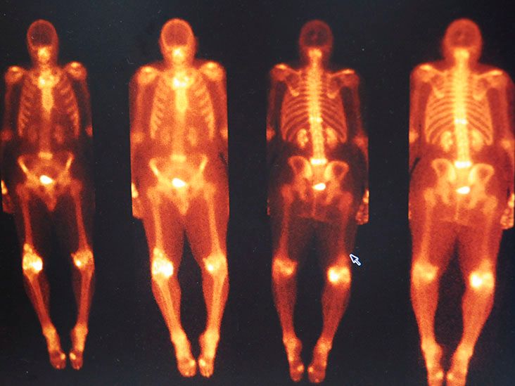
Cardiac amyloidosis used to be treated like the rare bird of heart disease: interesting, hard to spot, and usually discussed only after someone had already wandered through a maze of tests. But newer bone scan research is changing that picture. The big takeaway? Cardiac amyloidosis may still be uncommon in the general population, but among older adults, heart failure patients, and people referred for cardiac imaging, it is not nearly as rare as many once believed.
The phrase sounds intimidating, and, to be fair, it does not exactly roll off the tongue like “seasonal allergies.” Cardiac amyloidosis happens when abnormal protein deposits, called amyloid fibrils, build up in the heart muscle. Over time, those deposits can make the heart thick, stiff, and less able to fill or pump efficiently. The result can look a lot like common heart failure, especially heart failure with preserved ejection fraction, often shortened to HFpEF.
That overlap is one reason the condition is frequently missed. Patients may have shortness of breath, leg swelling, fatigue, dizziness, irregular heartbeat, or exercise intolerance. Those symptoms can be blamed on aging, high blood pressure, valve disease, kidney disease, or “getting out of shape.” The heart, unfortunately, does not send push notifications saying, “Amyloid deposits detected.” Clinicians have to suspect it.
What the Bone Scan Study Found
A large nuclear medicine bone scan study examined more than 17,000 scans from over 11,000 people and looked for cardiac uptake of a bone-seeking tracer called DPD. In this setting, uptake in the heart can be a clue to transthyretin cardiac amyloidosis, also known as ATTR cardiac amyloidosis or ATTR-CM.
The study found that about 3.3% of subjects had some degree of cardiac DPD uptake. That included about 1.8% with low-grade uptake and about 1.5% with grade 2 or 3 uptake, the stronger pattern more consistent with clinically meaningful cardiac amyloidosis. The prevalence was not evenly spread across everyone. Among people referred for non-cardiac reasons, the figure was roughly 1 in 50. Among cardiac referrals, it was closer to 1 in 5. In plain English: when doctors already had a heart-related reason to look, the scan found possible amyloid far more often.
The study also reported an important outcome signal. People with grade 2 or 3 uptake had a higher risk of heart failure hospitalization compared with those with no uptake. That does not mean every positive scan automatically predicts disaster, but it does mean this finding should not be shrugged off like a weird typo in a lab report.
So, How Common Is Cardiac Amyloidosis Really?
The most honest answer is: it depends on who you are looking at. In the broad public, cardiac amyloidosis remains relatively uncommon. But in certain groups, especially older adults with unexplained heart failure, thickened heart walls, atrial fibrillation, aortic stenosis, carpal tunnel syndrome, or spinal stenosis, the prevalence can rise sharply.
Studies of older patients with HFpEF and increased left ventricular wall thickness have found ATTR-CM in a meaningful minority of patients. In one community-based screening study, ATTR-CM was identified in more than 6% of screened patients with HFpEF and wall thickening, with higher rates in older men. Among male HFpEF patients in that screened group, the rate was around 10%. In the oldest age groups, prevalence climbed even higher.
Other research has suggested that ATTR cardiac amyloidosis may be present in a notable share of patients undergoing evaluation for severe aortic stenosis or transcatheter aortic valve replacement. That makes sense biologically and clinically: both conditions often affect older adults, both can cause shortness of breath and exercise limitation, and both can produce a heart that looks thick and stiff on imaging.
In other words, cardiac amyloidosis is not “common” in the same way high blood pressure is common. But it is common enough in the right clinical neighborhoods that doctors should stop acting surprised when it shows up. Think of it less like a unicorn and more like a raccoon in the attic: not in every house, but definitely worth checking when the clues are there.
Why Bone Scans Changed the Conversation
For decades, confirming cardiac amyloidosis often required a heart biopsy. A biopsy can be very useful, especially when the diagnosis is unclear, but it is invasive and not exactly anyone’s idea of a relaxing Tuesday. The rise of nuclear bone-avid tracer imaging has made it possible to diagnose many cases of ATTR-CM noninvasively when the scan is strongly positive and blood and urine testing rule out AL amyloidosis.
In the United States, clinicians commonly use a technetium-99m pyrophosphate scan, often called a PYP scan. In some other countries, DPD or HMDP tracers are used. These tracers were originally associated with bone imaging, but they can also bind in patterns that reveal ATTR amyloid deposits in the heart. That is the medical equivalent of buying a flashlight for the garage and discovering it also finds the raccoon.
What the Scan Grades Mean
Bone scan results are often described with visual grades. Grade 0 means no meaningful cardiac uptake. Grade 1 is low-grade or equivocal uptake and usually requires more evaluation. Grades 2 and 3 are stronger patterns and, when matched with the right clinical picture and negative monoclonal protein testing, can support a non-biopsy diagnosis of ATTR-CM.
Modern protocols also emphasize SPECT or SPECT/CT imaging, not just flat planar images. That matters because tracer activity can sometimes appear to be in the heart when it is actually in the blood pool, ribs, or nearby structures. A good scan is not just about taking pictures; it is about knowing exactly what is lighting up and why.
ATTR vs. AL: The Distinction That Really Matters
There are two major types of cardiac amyloidosis clinicians must separate quickly: AL amyloidosis and ATTR amyloidosis. AL amyloidosis is caused by abnormal light-chain proteins, usually related to a plasma cell disorder. It can progress rapidly and often needs urgent hematology-directed treatment. ATTR amyloidosis is caused by misfolded transthyretin protein, which is produced mainly by the liver. ATTR can be hereditary or wild-type, with wild-type ATTR more often affecting older adults, especially men.
This distinction is crucial because a positive bone scan alone is not enough to ignore AL amyloidosis. Before clinicians confidently call a scan ATTR-CM, they typically perform a monoclonal protein screen. That includes serum free light chain testing and serum and urine immunofixation. If those tests suggest a monoclonal protein, biopsy and specialized amyloid typing may be needed.
Why all the caution? Because treating ATTR like AL, or AL like ATTR, is like bringing a snow shovel to a beach cleanup. Both are tools. One is wildly wrong for the job.
Who Is Most Likely to Have Cardiac Amyloidosis?
Cardiac amyloidosis becomes more likely with age, especially ATTR-CM. Wild-type ATTR tends to appear in older adults and has a strong male predominance. Hereditary ATTR depends on specific TTR gene variants. One important variant, Val122Ile, is more common among Black Americans and people with West African ancestry, although not everyone who carries the variant develops cardiomyopathy.
Risk is also higher when certain clues cluster together. A patient with heart failure symptoms, preserved ejection fraction, thickened heart walls, atrial fibrillation, and a history of bilateral carpal tunnel syndrome should make clinicians pause. Add spinal stenosis, unexplained neuropathy, low blood pressure, kidney involvement, or a family history of cardiomyopathy, and the “maybe amyloidosis” light should get even brighter.
Common Red Flags Doctors Look For
Several findings can point toward cardiac amyloidosis. These include unexplained left ventricular wall thickening, heart failure with preserved ejection fraction, diastolic dysfunction, atrial fibrillation, conduction disease requiring a pacemaker, elevated cardiac biomarkers, low voltage on ECG despite a thick heart on echocardiogram, carpal tunnel syndrome, spinal stenosis, biceps tendon rupture, peripheral neuropathy, autonomic symptoms, and protein in the urine.
Some clues are more suggestive of AL amyloidosis, such as an enlarged tongue, easy bruising around the eyes, or certain bleeding abnormalities. ATTR has its own favorite side quests, including spinal stenosis and tendon or ligament problems. The trick is not to treat these clues as random trivia. Together, they may form a diagnostic breadcrumb trail.
Why the Disease Is Often Missed
Cardiac amyloidosis is underdiagnosed because it is excellent at wearing disguises. It can look like hypertensive heart disease. It can look like ordinary HFpEF. It can look like aortic stenosis, kidney-related fluid overload, atrial fibrillation, or simple aging. Many patients see several clinicians before the correct diagnosis appears.
Another reason is that the disease can progress slowly. Someone may have carpal tunnel surgery years before heart symptoms become obvious. Someone else may develop spinal stenosis, then atrial fibrillation, then mild shortness of breath, with each event treated as separate. Only later does someone connect the dots and say, “Wait a minute. These dots are suspiciously dot-shaped.”
There is also a historical bias. For years, cardiac amyloidosis was considered rare, hard to diagnose, and difficult to treat. That combination discouraged aggressive testing. Now that noninvasive imaging is widely available and disease-modifying treatments exist for ATTR-CM, early recognition matters much more.
What the Bone Scan Study Means for Patients
The study does not mean everyone needs a cardiac amyloidosis scan. Screening the entire population would not make sense. But it does support a more practical message: when older adults have unexplained heart failure, thickened heart walls, or compatible red flags, clinicians should consider cardiac amyloidosis earlier.
For patients, the message is not to self-diagnose after reading one article at midnight with a bowl of cereal nearby. The better move is to bring a pattern of symptoms and medical history to a clinician’s attention. For example, “I have heart failure with preserved ejection fraction, thickened heart walls, and I had carpal tunnel in both wrists” is a much more useful sentence than “The internet says my heart is full of protein confetti.”
What Happens After a Suspicious Scan?
If a bone scan suggests ATTR-CM, the next steps depend on the full clinical picture. Doctors generally confirm that AL amyloidosis has been ruled out with the appropriate blood and urine tests. If ATTR-CM is diagnosed, genetic testing is often recommended to distinguish hereditary ATTR from wild-type ATTR. That matters for family counseling and long-term care planning.
Treatment may include disease-modifying therapy, careful fluid management, rhythm monitoring, anticoagulation when needed, and coordinated care among cardiology, hematology, neurology, nephrology, and genetics specialists. Standard heart failure medications are not always tolerated in cardiac amyloidosis, especially when low blood pressure or autonomic dysfunction is present. This is one reason specialized care can make a major difference.
Why Prevalence Studies Are Not All the Same
Prevalence numbers can seem confusing because studies examine different groups. A study of all-comer bone scans is not the same as a study of hospitalized HFpEF patients. A community screening study is not the same as a referral-center study. A group of 90-year-old men with thickened heart walls is not the same as a group of healthy 45-year-olds.
That is why the answer to “How common is cardiac amyloidosis?” needs context. In younger people without symptoms, it is rare. In older adults with unexplained HFpEF and ventricular thickening, it becomes much more plausible. In selected cardiac referral groups, especially those already showing suspicious imaging or clinical clues, it can be surprisingly frequent.
The bone scan study is important because it shows that cardiac tracer uptake can be found incidentally in people undergoing scans for various reasons. It also shows that higher-grade uptake is not merely an imaging curiosity. It may carry prognostic weight, especially for heart failure outcomes.
The Bigger Public Health Message
Cardiac amyloidosis sits at the intersection of aging, heart failure, imaging, genetics, and specialty care. As the population ages and clinicians use better diagnostic tools, more cases will likely be found. Some of that rise reflects increased awareness rather than a true explosion in disease. Still, the practical result is the same: more patients can be diagnosed before advanced damage occurs.
Earlier diagnosis is valuable because modern ATTR-CM treatments aim to slow disease progression. They are not magic erasers for existing scar-like deposits and structural damage. This makes timing important. Finding the disease after years of decline is better than never finding it, but finding it before major functional loss is better still.
Experiences Related to Cardiac Amyloidosis and Bone Scan Prevalence
Real-world experiences around cardiac amyloidosis often share a familiar theme: the diagnosis arrives late, but the clues were hiding in plain sight. A typical patient story might begin with something ordinary, such as fatigue while walking uphill or swelling around the ankles after dinner. The person may be told they have heart failure with preserved ejection fraction, which is accurate but not always complete. HFpEF is a syndrome, not always the final explanation. It tells us the heart is struggling to fill properly, but it does not always say why.
Another common experience involves a long medical timeline. A patient may have had carpal tunnel syndrome in both wrists years earlier, then lumbar spinal stenosis, then atrial fibrillation, then shortness of breath. Each problem is real, and each may be treated separately. The wrist surgeon fixes the wrist. The spine specialist treats the back. The cardiologist manages the rhythm. But unless someone steps back and sees the pattern, cardiac amyloidosis can remain the uninvited guest standing quietly in the corner.
For many people, the bone scan is the turning point. Compared with a biopsy, a PYP scan or similar nuclear scan can feel almost anticlimactic. The patient receives a small injection of tracer, waits, lies still for images, and then goes home. There is no dramatic hospital scene, no glowing superhero origin story, and usually no special preparation. Yet the scan can provide a powerful clue when interpreted correctly with SPECT imaging and laboratory testing.
Patients often describe mixed emotions after diagnosis. There may be relief because months or years of vague symptoms finally have a name. There may also be frustration: “Why did it take so long?” That frustration is understandable. Cardiac amyloidosis has historically been underrecognized, and many clinicians were trained during an era when it was considered exceptionally rare. The new prevalence data challenges that older mindset.
Families can also be affected, especially when hereditary ATTR is found. Genetic testing may raise questions for siblings, children, and extended relatives. This is where genetic counseling becomes important. A gene variant is not the same thing as a guaranteed future illness, but it may help relatives make informed decisions about monitoring and evaluation.
Clinicians have their own experience curve. Cardiologists, nuclear medicine physicians, primary care doctors, hematologists, and neurologists are learning to recognize the condition earlier. A bone scan study showing meaningful prevalence helps shift cardiac amyloidosis from “rare zebra” to “possible diagnosis when the hoofbeats sound strange.” The goal is not to scan everyone. The goal is to scan the right people sooner.
For patients living with ATTR-CM, day-to-day experience may involve managing fluid balance, monitoring weight, adjusting diuretics, watching for rhythm problems, staying active within safe limits, and keeping regular appointments. The emotional work matters too. A diagnosis can feel heavy, but it can also open the door to targeted care, clearer expectations, and fewer unanswered questions.
The most practical lesson from these experiences is simple: patterns matter. One symptom may be ordinary. Several clues together may deserve a second look. If an older adult has unexplained HFpEF, thickened heart walls, atrial fibrillation, carpal tunnel history, spinal stenosis, neuropathy, or a family history of amyloidosis, asking about ATTR-CM is reasonable. Not panicked. Not dramatic. Just reasonable.
Conclusion
Cardiac amyloidosis is not as rare as yesterday’s medical folklore suggested, especially among older adults with heart failure symptoms and thickened heart walls. The bone scan study adds weight to that message by showing that cardiac tracer uptake appears in a measurable share of scanned patients and is more common in cardiac referrals. Stronger uptake also appears linked with worse heart failure outcomes, making it a finding worth taking seriously.
The best interpretation is balanced: cardiac amyloidosis is not common enough to screen every person, but it is common enough to consider in the right clinical setting. Bone-avid tracer imaging, combined with careful blood and urine testing to rule out AL amyloidosis, has transformed diagnosis. For patients, that means fewer invasive biopsies in many cases and a better chance of identifying ATTR-CM while treatment can still help preserve function.
Note: This article is for educational purposes only and should not replace professional medical evaluation. Anyone with symptoms such as unexplained shortness of breath, swelling, fainting, irregular heartbeat, or suspected heart failure should speak with a qualified healthcare professional.